The eye
Our eyes are among the most impressive organs in our body. With the ability to filter light, translate images, and communicate with the brain, few body parts can handle and process as much stimuli and information as our eyes. In a way far superior to any electronic device, our eyes are capable of interpreting an incredible amount of sensory details in virtually no time at all, giving us the ability to perceive the world we live in. Plus, they’re beautiful to gaze into. As with any complex bodily structure, however, sometimes, things can go wrong. Around ninety percent of the population has imperfect vision and over sixty percent need some sort of corrective lens or, in some cases, surgery, to maintain an acceptable level of vision.
The parts of the eye
The eyeball is set within the ocular sockets of the skull and is connected to the brain via a passage of nerves. The front of the eye is covered by a clear lens, allowing light and images to filter through. The eye itself is made up of numerous structures, tissues and cells.
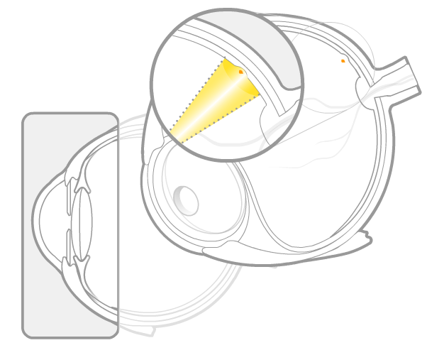
Cornea
The cornea is the thin, clear layer on the front of the eye covering the pupil, iris and lens. The cornea assists in refracting, or bending, light in a way that focuses the light onto the retina.
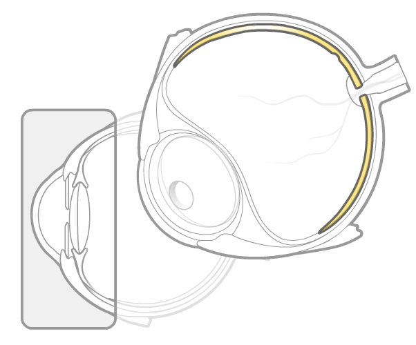
Sclera
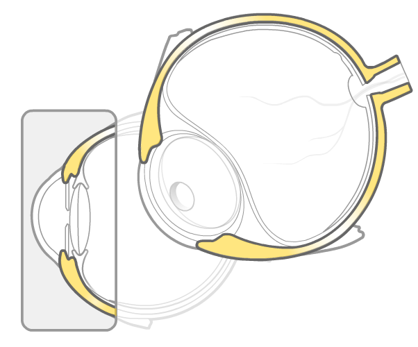
The sclera is the opaque, white coating of the eyeball composed of elastic fiber and collagen. It serves as a protective shell for the eye.
Iris
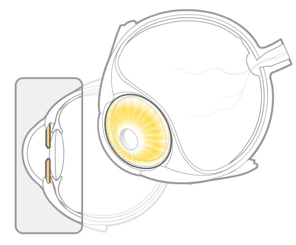
The iris is the colored, doughnut-shaped part of the eye covered by the cornea and surrounded by the sclera. The iris opens and closes to change the size of the pupil, controlling the amount of light permitted to enter the eye.
Pupil
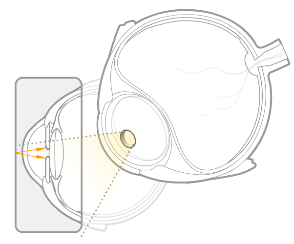
The pupil is the dark hole in the center of the iris. The pupil is expanded and contracted by the iris, allowing in more light in dark surroundings and less when bright to control how much light enters the eye.
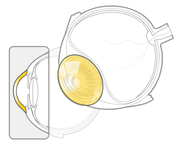
Crystalline lens
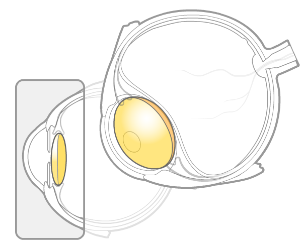
The crystalline lens is a structure made of transparent tissue behind the cornea and pupil that acts like a magnifying glass. When we focus our eyes, the lens changes shape to allow us to focus at different distances. Over time, the lens hardens, making it less flexible, a main contributing factor to the need for bifocals or reading glasses.
Conjunctiva
The conjunctiva is the thin lining under the eyelid and over the sclera. It is made up of epithelial cells, or skin cells, and goblet cells. It is responsible for lubricating the eye and is part of the eye’s protective immune response. The red veins over the sclera are the blood vessels located within the conjunctiva so when you wake up with bloodshot eyes you can clench your fists and the heavens and yell, “Arrrrrg! Stupid conjunctiva!”
Aqueous humor
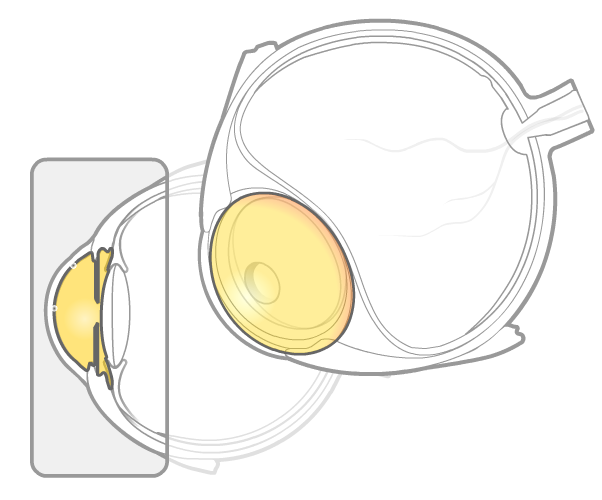
The watery fluid filling the space behind the cornea and forward of the crystalline lens. Its main function is to provide nutrients to the front portion of the eyeball.
Retina
The retina is the layer of nerve cells lining the inside of the eye. The retina is very sensitive to light and contains the receptor cells, called rods and cones that convert light signals into electrical impulses that are transmitted to the brain. Rod cells are used to see in dim light while cone cells assist in the perception of colors.
Macula
The macula is the area in the center of the retina responsible for helping us see details and colors. The center of the macula is the fovea, the area containing the highest concentration of cone cells.
Optic nerve
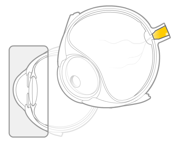
The optic nerve is the large bundle of nerves that connects the eye to the brain that transmits the impulses from rod and cone cells.
Vitreous humor
The vitreous humor is the clear, gelatinous structure located behind the crystalline lens and taking up most of the volume inside the eye. The primary purpose of the vitreous humor is to keep the retina against the inner wall of the eyeball and, frankly, we think it does a fine job of it.
Choroid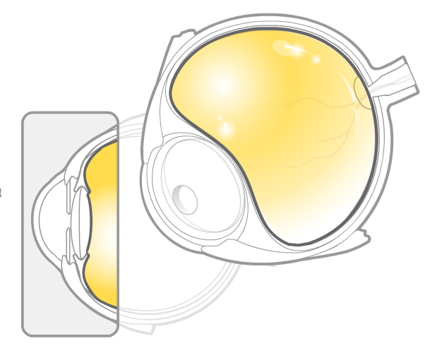
The choroid is the middle layer of the eyeball, between the sclera and the retina. Its primary purpose is to supply nutrients to the retina.
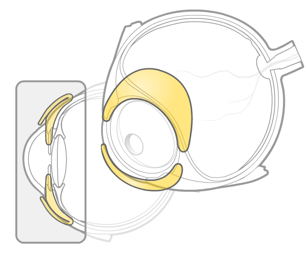
Ciliary body
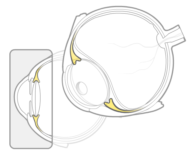
The ciliary body is the extension of the choroid that connects to the iris and the lens, via thin nerve fibers called zonules. It controls the movement of the crystalline lens and produces the aqueous humor (not funny).